- HOME
- Technical Information
- Commissioned Analysis and Research
- Transmission Electron Microscope : TEM
Transmission Electron Microscope : TEM
Principle
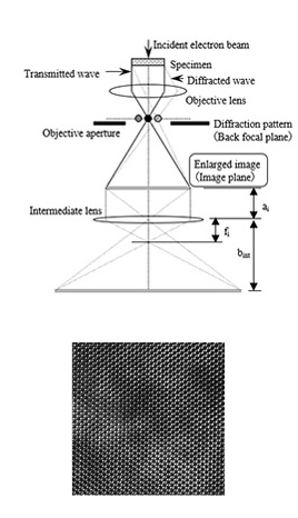
We observe the image obtained using the transmitted electrons by irradiating a thin sample with an electron beam accelerated to tens to hundreds of kV. Microscopic images related to the crystal structure can be obtained using transmission electrons and diffracted electrons. TEM specimen preparation methods include the ultra-thin section method, Ar-ion milling method, and FIB method are used depending on the type of material and other requirements.
General layout of a TEM
describing the path of electron beam
in a TEM
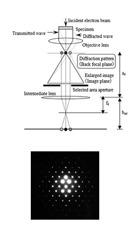
A ray diagram for
the diffraction mechanism in TEM
electron diffraction pattern