- HOME
- Technical Information
- Commissioned Analysis and Research
- Scanning Electron Microscope : SEM
Scanning Electron Microscope : SEM
Principle
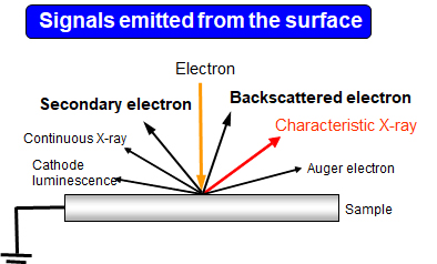
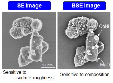
In SEM, the electron beam is focused by magnetic field lens and irradiate onto the sample. The incident electron beam is scanned over the sample, and the image is obtained by detecting the secondary electrons (SE) and the backscattered electrons (BSE) emitted from the sample. The contrast of SE image mainly depends on the surface roughness, and that of BSE image mainly depends on the composition. In addition, it is equipped with EDX (Energy Dispersive X-ray Spectroscopy) detector, which enables elemental analysis by detecting characteristic X-rays.